G-biosciences LDH细胞毒性检测试剂盒

产品名称:G-biosciences LDH细胞毒性检测试剂盒

产品货号:  786-324

产品规格:200 Assays

产品品牌:G-biosciences


产品简介:

G-biosciences LDH细胞毒性检测试剂盒通过比色法,可测量稳定的细胞溶质乳酸脱氢酶 (LDH),该酶从受损细胞中释放出来,是细胞毒性和细胞溶解的生物标志物。释放的 LDH 是通过偶联酶促反应测量的,该反应导致四唑盐(碘硝基四唑 (INT))通过黄递酶转化为红色甲臜。LDH 活性被确定为 NADH 氧化或 INT 在定义的时间段内的还原。所得甲臜在 492nm 处吸收最大,可在 490nm 处定量测量。CytoScan™ LDH 细胞毒性检测试剂盒可用于不同细胞类型,用于检测细胞介导的细胞毒性以及化学物质和其他测试化合物介导的细胞毒性。

产品特点

1.定量测量LDH释放

2.比色法

3.高通量筛选的理想选择

产品应用:

用于检测细胞毒性、细胞死亡、细胞活力或细胞增殖

产品组分:

Cytoscan™ Substrate Mix (lyophilizate) 5 1

Cytoscan™-LDH Assay Buffer 5 x 0.6ml 1 x 0.6ml

Cytoscan™-LDH Lysis Buffer [10X] 12ml 2.5ml

Cytoscan™-LDH Stop Solution 60ml 12ml

储存条件:

常温运输。

存储Cytoscan™Substrate Mix和Assay缓冲区在-20°C。LDH阳性对照,10X裂解缓冲液和终止液可在4°C。

效期1个月。


产品订购信息:

货号               名称                           规格

786-324 LDH细胞毒性检测试剂盒    200 Assays

产品名称:G-biosciences LDH细胞毒性检测试剂盒

产品货号:  786-324

关于公司:

Geno Technology, Inc.成立的愿景是简化生命科学研究。我们的主要目标是针对蛋白质研究的关键技术,并找到负担得起的方法来简化和改进这些技术。多年来,Geno技术公司已经扩展到其他研究领域,但我们的主要目标仍然是蛋白质,我们的主要目标“简化和改进这些技术”仍然是正确的。2010年,我们标志性的吉祥物“蛋白质人”和G-Biosciences品牌(Geno Technology, Inc.的注册商标)的演变进一步强化了这一信息。

CytoScan 乳酸脱氢酶细胞毒性检测试剂盒

产品名称:CytoScan 乳酸脱氢酶细胞毒性检测试剂盒

产品货号:786-324

英文名称:CytoScan™ LDH Cytotoxicity Assay

产品规格:200 Assays

产品品牌:G-biosciences


产品简介:

一种可靠的比色法,可测量稳定的细胞溶质乳酸脱氢酶 (LDH),该酶从受损细胞中释放出来,是细胞毒性和细胞溶解的生物标志物。释放的 LDH 是通过偶联酶促反应测量的,该反应导致四唑盐(碘硝基四唑 (INT))通过黄递酶转化为红色甲臜。LDH 活性被确定为 NADH 氧化或 INT 在定义的时间段内的还原。所得甲臜在 492nm 处吸收最大,可在 490nm 处定量测量。CytoScan™ LDH 细胞毒性检测试剂盒可用于不同细胞类型,用于检测细胞介导的细胞毒性以及化学物质和其他测试化合物介导的细胞毒性。

51 Cr 释放细胞毒性测定的替代 方法。该试剂盒结合了放射性同位素释放测定的可靠性和评估简单的优点,以及准确性和避免放射性的便利性。


产品特点:

定量测量LDH释放

比色法

高通量筛选的理想选择


产品应用:

用于检测细胞毒性、细胞死亡、细胞活力或细胞增殖

对于来自培养细胞(贴壁或悬浮)的无细胞上清液


产品订购信息:

货号               名称                                          规格

786-200A CasPASE1, 4, 5assay含Ac-WEHD-AFC底物  50 Assays


产品组分:

Cytoscan™ Substrate Mix (lyophilizate) 5 1

Cytoscan™-LDH Assay Buffer 5 x 0.6ml 1 x 0.6ml

Cytoscan™-LDH Lysis Buffer [10X] 12ml 2.5ml

Cytoscan™-LDH Stop Solution 60ml 12ml

LDH Positive Control 30µl 6µl


储存运输;

该套件在环境温度下运输。存储Cytoscan™Substrate Mix和Assay缓冲区在-20°C。LDH阳性对照,10X裂解缓冲液和终止液可在4°C。如果按照推荐的方式储存,该试剂盒可以稳定12个月。

产品名称:CytoScan 乳酸脱氢酶细胞毒性检测试剂盒

产品货号:786-324

关于公司:

Geno Technology, Inc. 的成立旨在简化生命科学研究。我们的主要目标是针对蛋白质研究的关键技术,并找到经济实惠的方法来简化和改进这些技术。多年来,Geno Technology, Inc. 已经扩展到其他研究领域,但我们的主要目标仍然是蛋白质,我们的主要目标“简化和改进这些技术”仍然成立。这一信息在 2010 年随着我们标志性吉祥物“蛋白质人”和 G-Biosciences 品牌(Geno Technology, Inc. 的注册商标)的演变而得到加强。

G-Bioscience 的团队检查了特定实验技术的各个方面,从蛋白质提取到蛋白质纯化和检测,以确定我们可以针对改进的关键领域。因此,我们现在生产各种产品,从浓度和尺寸方便的简单缓冲液到定制设备,例如 Tube-O-Dialyzer,可增强和简化关键技术。我们关注的关键领域包括但不限于蛋白质测定、样品制备、电泳、蛋白质印迹、质谱、测定开发、抗体生产和蛋白质纯化。

G-Biosciences 现在为蛋白质和生命科学研究市场提供数千种产品,所有产品的设计都是为了让研究人员的实验室时间更简单、更实惠。

CK12/Cytotoxicity LDH Assay Kit-WST-LDH检测试剂盒

LDH检测试剂盒—Cytotoxicity LDH Assay Kit-WST,Cytotoxicity LDH Assay Kit-WST,Cytotoxicity LDH Assay Kit-WST®是通过测定细胞释放到培养基中的乳酸脱氢酶 (LDH) 活性进而测定细胞损害的试剂盒。LDH是存在于细胞质的一种酶,当细胞膜受到损伤时,LDH会释放到培养基中。由于释放出的LDH稳定,检 测培养基中LDH量可以作为测定死细胞和受损细胞数量的指标。

货号:CK12
乳酸脱氢酶(LDH)检测试剂盒
Cytotoxicity LDH Assay Kit-WST
储存条件:0-5度保存,避光
运输条件:室温

特点:

  1. 不与细胞内LDH反应,可直接检测(一步法)或取上清液检测(低损伤法)。
  2. 更适合Car-T、ADCC、CDC等效靶杀伤实验。
  3. Working Solution配制后,可稳定保存6个月以上。
  4. 试剂盒稳定性高,数据重复性好。
  5. 搭配CCK-8检测,细胞毒性双指标认证。

选择规格:100 tests,500 tests,2000 tests

关联产品:

NO.1. Cell Counting Kit-8 细胞增殖毒性检测

NO.2. Annexin V, FITC Apoptosis Detection Kit 细胞凋亡检测

NO.3. Caspase-3 Assay Kit -Colorimetric 细胞凋亡检测

NO.4. Biotin Labeling Kit – NH2 生物素抗体标记-氨基

NO.5. Cell Counting Kit-Luminescence 细胞增殖/毒性检测

试剂盒内含

100 tests Dye Mixture
·Assay Buffer
-Lysis Buffer
Stop Solution
×1
11 ml×1
1.1 ml×1
5.5 ml×1
500 tests · Dye Mixture
-Assay Buffer
-Lysis Buffer
-Stop Solution
x1
55ml×1
5.5 ml×1
27.5 ml×1
2000 tests -Dye Mixture
·Assay Buffer
Lysis Buffer
-Stop Solution
x4
55ml×4
5.5 ml×4
27.5 ml×4

产品概述

Cytotoxicity LDH Assay Kit-WST®是通过测定细胞释放到培养基中的乳酸脱氢酶 (LDH) 活性进而测定细胞损害的试剂盒。LDH是存在于细胞质的一种酶,当细胞膜受到损伤时,LDH会释放到培养基中。由于释放出的LDH稳定,检 测培养基中LDH量可以作为测定死细胞和受损细胞数量的指标。本试剂盒可以测定受损细胞所释放出的LDH,其原 理是LDH催化乳酸生成丙酮酸和NADH,NADH通过电子载体可将水溶性四唑盐WST® (无色) 还原成甲臜产物 (橙色), WST®甲臜产物的吸光度与LDH量呈正比,利用此原理可以测定死细胞和受损细胞的数量。

本试剂盒中试剂不会和活细胞反应,而且对细胞无损害,可以直接添加到含有细胞的培养基中检测 (一步法)。也可以分离细胞后,加到培养基中检测 (低损伤法,分离的细胞可以做其他实验)。本试剂盒采用稳定性高的WST®,配制后的溶液可以长时间保存,不需要现配现用,适用于高通量筛选。

原理

Cytotoxicity LDH Assay Kit-WST®是通过测定细胞释放到培养基中的乳酸脱氢酶(LDH)活性进而测定细胞损害的试剂盒。LDH(乳酸脱氢酶)是存在于细胞质的一种酶,当细胞膜受到损伤时,LDH 会释放到培养基中。由于释放出的LDH 稳定,检测培养基中LDH 的量可以作为测定死细胞和受损细胞数量的指标。

本试剂盒可以测定受损细胞所释放出的LDH,其原理是LDH 催化乳酸生成丙酮酸和NADH,NADH 通过电子载体可将水溶性四唑盐WST®(无色)还原成甲臜产物(橙色),WST®甲臜产物的吸光度与LDH 的量呈正比,利用此原理可以测定死细胞和受损细胞的数量。

本试剂盒中试剂不会和活细胞反应,而且对细胞无损害,可以直接添加到含有细胞的培养基中检测(一步法)。也可以分离细胞后,加到培养基中检测(低损伤法,分离的细胞可以做其他实验)。本试剂盒采用稳定性高的WST®,配置后的溶液可以长时间保存6个月,不需要现配现用,适用于高通量筛选。


产品特点

-双重选择:有2种检测方法(一步法、低损伤法)

-操作简单:不需要分离死细胞和活细胞,就可检测死细胞的LDH *一步法

-仪器常用:采用常规酶标仪检测

-稳定方便:Working Solution可冷藏保存6个月,不需要现配现用

-双重验证:配合使用CCK-8可获得死细胞和活细胞的结果

操作示意图

本试剂盒不与细胞内的LDH反应,因此可选择两种检测方法。

1、一步法,直接添加的培养基中检测(左)

2、低损伤法,取上清液检测(右)

工作液稳定性

配制后的工作液,可冷藏保存6个月,无需现配现用

CCK-8与LDH

Cell Counting Kit-8 与Cytotoxicity LDH Assay Kit-WST®同时检测细胞毒性

检测药物: Mitomycin C
细胞: HeLa
培养基: MEM, 10% FBS
孵育条件: 37℃, 5% CO2, 48小时
检测波长: Cell Counting Kit-8 (450 nm), Cytotoxicity LDH Assay Kit-WST® (490 nm)

使用Mitomycin C 检测检测HeLa细胞毒性。通过使用Cell Counting Kit-8和Cytotoxicity LDH Assay Kit-WST [货号: CK12]两种方法检测。Cell Counting Kit-8以细胞活性为指标,Cytotoxicity LDH Assay Kit-WST以游离的LDH为指标,实验证实两种指标检测存在差异性。二者联用,能更全面的表征细胞毒性。

本文为LDH法检测细胞毒性的相关研究介绍。

实验例

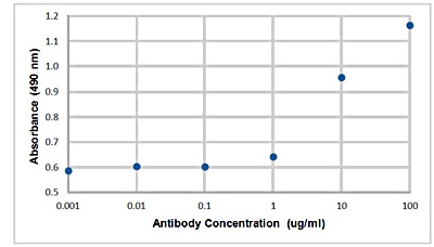
RAJI细胞CDC实验

实验证实:吸光度O.D.值与抗体浓度成正相关,因此确认CD20抗体与细胞结合而激活细胞损伤
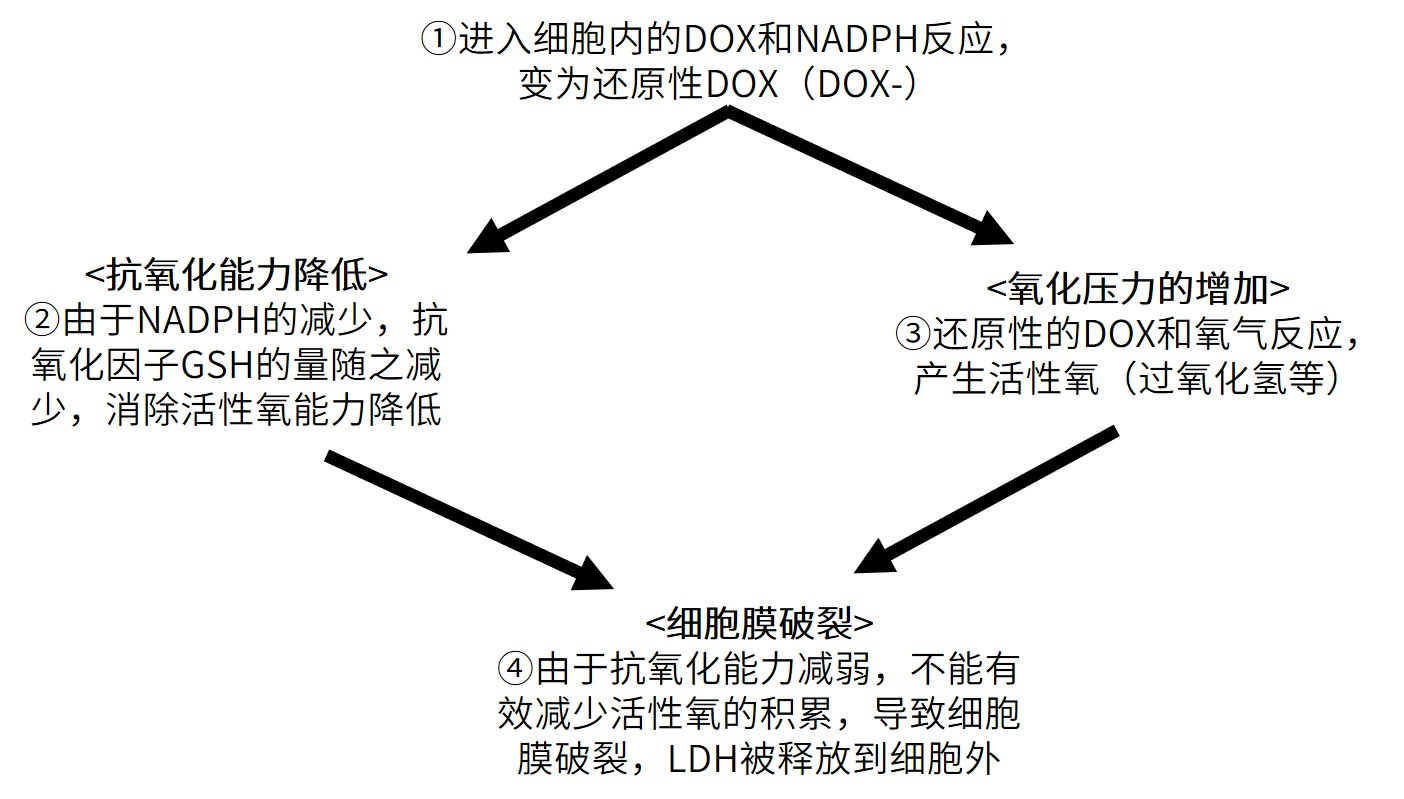
抗癌药阿霉素(DOX)的细胞毒性机制

近年来,与抗氧化剂能力相关的指标(NADP + / NADPH,GSSG / GSH等)与细胞毒性评估一起被测量的案例越来越多。

在本文中,我们将以DOX为例,其中抗氧化能力的下降与细胞毒性的增加有关。

常见问题Q&A

Q1:可以使用490 nm以外的滤光片吗?

A1:可以使用450 nm滤光片,吸光度会高于490 nm。

Q2:细胞毒性检测时,Cytotoxicity LDH Assay Kit-WST和Cell Counting Kit-8法结果一致吗?

A2:二者测定的原理和指标不同,建议同时检测。可更好说明问题。

Cell Counting Kit-8:检测细胞活性

Q3:LD50差异性过大,如何处理?

A3:细胞状态不同,对药物的耐受性也会有很大变化,最终影响LD50的数值。请尽可能使用相同条件下处理、相同代数的细胞。在准备药物稀释液时,可降低稀释梯度倍数,从而得到更准确的LD50。

Q4:这个试剂盒能检测LDH的酶活吗?

A4:本产品通过LDH活性检测细胞毒性,不能检测如:LDH1, LDH2, LDH3的活性。

Q5:LDH本身的稳定性如何?

A5:文献报道,培养基中LDH活性的1天内降低<5%(文献1),也可冷藏1周左右(文献2)。具体可因实验条件不同存在差异。

文献1:

A. Wagner, A. Marc, JM. Engasser, A. Einsele, “The use of lactate dehydrogenase (LDH) release kinetics for the evaluation of death and growth of mammalian cells in perfusion reactors.”, Biotechnol Bioeng. 1992; 39, (3), :320..

文献2:

M. Allen, P. Millett, E. Dawes, N. Rushton., “Lactate dehydrogenase activity as a rapid and sensitive test for the quantification of cell numbers in vitro.”, Clin Mater. 1994; 16, (4), 189.

Q6:Cytotoxicity LDH Assay Kit-WST带LDH标准品吗?

A6:因为是检测细胞毒性使用,不带标准品。

Q7:试剂盒中的Stop solution添加后需立即检测吗?

A7:Stop solution添加后可避光保存3小时。

Q8:显色背景过高,有什么原因?

A8:考虑以下原因。

培养基中血清中的乳酸脱氢酶升高了背景。请用此培养基配制小于5%的血清或血清培养基。

如果培养基中含有待测物质具有强还原性,会与WST®显色。因此可预实验时单独检测判断。

(一般情况下,培养基如DMEM、RPMI、F-12,通常不含还原性物质

参考文献

1.Antitumor Effect by Hydroxyapatite Nanospheres: Activation of Mitochondria-Dependent Apoptosis and Ne…

ACS Nano,2018,12(8)7838-7854

2.Magnetic Nano-Clusters Armed with Responsive PD-1 Antibody

3.Synergistically Improved Adoptive T C….ACS Nano,2019, 13(2)1469-1478

4.Layer by layer cell coating technique using extracellular matrix facilitates rapid fabrication and fu….Biomaterials,2018,160,82-91

5.Oxidative stress–dependent phosphorylation activates ZNRF1 to induce neuronal/axonal degeneration

J.Cell Biol.,2015,211(4)881-896

6.Protein carbamylation exacerbates vascular calcification.Kidney International ,2018,94(1),72-90

7.In vitro cellular behaviors and toxicity assays of small-sized fluorescent silicon nanoparticles.Nanoscale,2017, 9(22),7602-7611

8.Pathological Endogenous a-Synuclein Accumulation in Oligodendrocyte Precursor Cells Potentially Induc…

Stem Cell Reports,2018,10(2),356-365

9.Biocompatible carbon nanotube fiber for implantable supercapacitors.Carbon,2017,122,162-167

10.Lower Expression of MicroRNA-155 Contributes to Dysfunction of Natural Killer Cells in Patients with ….Frontiers in Immunology,2017,8,1173

11.Melatonin Mediates Protective Effects against Kainic Acid-Induced Neuronal Death through Safeguarding….Frontiers in Immunology,2017,10,49

ProSpec-FGF 9 Human

  • Description
  • FGF 9 Human

  • Fibroblast Growth Factor-9 Human Recombinant
  • CYT-415

Catalogue number

CYT-415

Synonyms

GAF , HBGF-9, MGC119914, MGC119915, FGF-9.

Introduction

The human FGF-9 cDNA encodes a 208 amino acid residue protein that contains a single, potential N-linked glycosylation site. The native protein is glycosylated and is efficiently secreted after synthesis, although FGF -9 lacks a typical secretion signal. Rat and mouse FGF-9 show a very high homology to human FGF-9. The transcripts for FGF-9 have been found in brain and in kidney tissue. Fibroblast Growth Factor-9 is a member of the fibroblast growth factor family. FGF family members possess broad mitogenic and cell survival activities, and are involved in a variety of biological processes, including embryonic development, cell growth, morphogenesis, tissue repair, tumor growth and invasion. FGF9 was isolated as a secreted factor that exhibits a growth-stimulating effect on cultured glial cells. In nervous system, this protein is produced mainly by neurons and may be important for glial cell development. Expression of the mouse homolog of this gene was found to be dependent on Sonic hedgehog signaling. Mice lacking the homolog gene displayed a male-to-female sex reversal phenotype, which suggested a role in testicular embryogenesis Fibroblast Growth Factor 9 may have a role in glial cell growth and differentiation during development, gliosis during repair and regeneration of brain tissue after damage, differentiation and survival of neuronal cells, and growth stimulation of glial tumors.

Description

Fibroblast Growth Factor-9 Human Recombinant produced in E.coli is a single, non-glycosylated, polypeptide chain containing 207 amino acids and having a molecular mass of 23.4 kDa. The FGF-9 is purified by proprietary chromatographic techniques.

Source

Escherichia coli.

Physical Appearance

Sterile Filtered white lyophilized powder.

Formulation

The sterile protein powder is lyophilized from 1mg/ml solution containing 1xPBS.

Solubility

It is recommended to reconstitute the lyophilized Fibroblast Growth Factor-9 Human Recombinant sterile 18M-cm H2O not less than 100µg/ml, which can then be further diluted to other aqueous solutions.

Stability

Lyophilized Fibroblast Growth Factor 9 Human Recombinant although stable at room temperature for 3 weeks, should be stored desiccated below -18°C. Upon reconstitution FGF-9 should be stored at 4°C between 2-7 days and for future use below -18°C.
For long term storage it is recommended to add a carrier protein .
Please prevent freeze-thaw cycles.

Purity

Greater than 95.0% as determined by RP-HPLC and -PAGE.

Amino acid sequence

APLGEVGNYF GVQDAVPFGN VPVLPVDSPV LLSDHLGQSE AGGLPRGPAV
TDLDHLKGIL RRRQLYCRTG FHLEIFPNGT IQGTRKDHSR FGILEFISIA
VGLVSIRGVD SGLYLGMNEK GELYGSEKLT QECVFREQFE ENWYNTYSSN
LYKHVDTGRR YYVALNKDGT PREGTRTKRH QKFTHFLPRP VDPDKVPELY
KDILSQS.

Biological Activity

The ED50, calculated by the dose-dependant proliferation of BAF3 cells expressing FGF receptors is <0.5ng/ml, corresponding to a specific activity of 2,000,000 Units/mg.

Usage

ProSpec’s products are furnished for LABORATORY RESEARCH USE ONLY.They may not be used as drugs,agricultural or pesticidal products, food additives or household chemicals.

Related Products

  • FGF 9 Rat
  • FGF 9 Mouse

ProSpec-FGF 9 Rat

  • Description
  • FGF 9 Rat

  • Fibroblast Growth Factor-9 Rat Recombinant
  • CYT-558

Catalogue number

CYT-558

Synonyms

GAF , HBGF-9, MGC119914, MGC119915, FGF-9.

Introduction

Rat and mouse FGF-9 show a very high homology to human FGF-9. The transcripts for FGF-9 have been found in brain and in kidney tissue. Fibroblast Growth Factor-9 is a member of the fibroblast growth factor family. FGF family members possess broad mitogenic and cell survival activities, and are involved in a variety of biological processes, including embryonic development, cell growth, morphogenesis, tissue repair, tumor growth and invasion. FGF9 was isolated as a secreted factor that exhibits a growth-stimulating effect on cultured glial cells. In nervous system, this protein is produced mainly by neurons and may be important for glial cell development. Expression of the mouse homolog of this gene was found to be dependent on Sonic hedgehog signaling. Mice lacking the homolog gene displayed a male-to-female sex reversal phenotype, which suggested a role in testicular embryogenesis Fibroblast Growth Factor 9 may have a role in glial cell growth and differentiation during development, gliosis during repair and regeneration of brain tissue after damage, differentiation and survival of neuronal cells, and growth stimulation of glial tumors.

Description

Rat FGF9 Recombinant produced in E.Coli is a single, non-glycosylated, polypeptide chain containing 207 amino acids and having a molecular mass of 23.3kDa.
The FGF-9 Mouse Recombinant is purified by proprietary chromatographic techniques.

Source

Escherichia Coli.

Physical Appearance

Sterile Filtered White lyophilized powder.

Formulation

The FGF-9 was lyophilized from a concentrated sterile solution containing 10mM NaP, pH-7.5 &, 75mM Ammonium Sulfate.

Solubility

It is recommended to reconstitute the lyophilized Rat FGF-9 in sterile 18MΩ-cm H2O not less than 100µg/ml, which can then be further diluted to other aqueous solutions.

Stability

Lyophilized Rat Fibroblast Growth Factor-9 although stable at room temperature for 3 weeks, should be stored desiccated below -18°C. Upon reconstitution FGF9 Rat Recombinant should be stored at 4°C between 2-7 days and for future use below -18°C.
For long term storage it is recommended to add a carrier protein .
Please prevent freeze-thaw cycles.

Purity

Greater than 95.0% as determined by:
Analysis by RP-HPLC.
Analysis by -PAGE.

Amino acid sequence

MPLGEVGSYFG VQDAVPFGNV PVLPVDSPVL LNDHLGQSEA GGLPRGPAVT DLDHLKGILR RRQLYCRTGF HLEIFPNGTI QGTRKDHSRF GILEFISIAV GLVSIRGVDS GLYLGMNEKG ELYGSEKLTQ ECVFREQFEE NWYNTYSSNL YKHVDTGRRY YVALNKDGTP REGTRTKRHQ KFTHFLPRPV DPDKVPELYK DILSQS.

Biological Activity

The ED50, calculated by the dose-dependant proliferation of BAF3 cells expressing FGF receptors is <0.5 ng/ml, corresponding to a specific activity of 2,000,000 Units/mg.

Usage

ProSpec’s products are furnished for LABORATORY RESEARCH USE ONLY. The product may not be used as drugs, agricultural or pesticidal products, food additives or household chemicals.

Related Products

  • FGF 9 Mouse
  • FGF 9 Human

ProSpec-FGFBP Human

  • Description
  • FGFBP Human

  • Fibroblast Growth Factor Binding Protein 1 Human Recombinant
  • CYT-860

Catalogue number

CYT-860

Synonyms

Fibroblast Growth Factor Binding Protein 1, FGFBP, HBP17, 17 KDa Heparin-Binding Growth Factor-Binding Protein, 17 KDa HBGF-Binding Protein, FGF-Binding Protein 1, FGF-BP1, FGFBP-1, FGF-BP, Heparin-Binding Growth Factor Binding Protein, Fibroblast Growth Factor-Binding Protein 1, Fibroblast growth factor-binding protein 1.

Introduction

Fibroblast Growth Factor Binding Protein 1, also known as FGFBP1 is a secreted fibroblast growth factor carrier protein. FGFBP1 plays a vital part in cell proliferation, differentiation and migration by binding to fibroblast growth factors and potentiating their biological effects on target cells. In addition, FGFBP1 also takes part in tumor growth as an angiogenic switch molecule, furthermore an expression of FGFBP1 has been associated with more than a few types of cancer as well as pancreatic and colorectal adenocarcinoma.

Description

FGFBP Human Recombinant produced in E.Coli is a single, non-glycosylated polypeptide chain containing 234 amino acids and having a molecular mass of 26.2kDa.

FGFBP is fused to a 23 amino acid His-tag at N-terminus & purified by proprietary chromatographic techniques.

Source

Escherichia Coli.

Physical Appearance

Sterile Filtered colorless clear solution.

Formulation

FGFBP protein solution containing Phosphate buffered saline , 10% glycerol and 1mM DTT.

Stability

Store at 4°C if entire vial will be used within 2-4 weeks.
Store, frozen at -20°C for longer periods of time.
For long term storage it is recommended to add a carrier protein .
Please avoid freeze thaw cycles.

Purity

Greater than 85.0% as determined by -PAGE.

Amino acid sequence

MGSSHHHHHH SSGLVPRGSH MGSKKKVKNG LHSKVVSEQK DTLGNTQIKQ KSRPGNKGKF VTKDQANCRW AATEQEEGIS LKVECTQLDH EFSCVFAGNP TSCLKLKDER VYWKQVARNL RSQKDICRYS KTAVKTRVCR KDFPESSLKL VSSTLFGNTK PRKEKTEMSP REHIKGKETT PSSLAVTQTM ATKAPECVED PDMANQRKTA LEFCGETWSS LCTFFLSIVQ DTSC.

Usage

ProSpec’s products are furnished for LABORATORY RESEARCH USE ONLY. They may not be used as drugs,agricultural or pesticidal products, food additives or household chemicals.

ProSpec-HGF A Human

  • Description
  • HGF A Human

  • Hepatocyte Growth Factor A Chain Human Recombinant
  • CYT-664

Catalogue number

CYT-664

Synonyms

Scatter Factor, SF, Hepatopoietin, HPTA, HGF, HGFB, F-TCF, DFNB39, Hepatocyte growth factor, Hepatopoeitin-A, Hepatocyte growth factor alpha chain.

Introduction

Hepatocyte Growth Factor is a multifunctional growth factor which regulates both cell growth and cell motility. It exerts a strong mitogenic effect on hepatocytes and primary epithelial cells. HGF synergizes with Interleukin-3 and GM-CSF to stimulate colony formation of hematopoietic progenitor cells in vitro and may, therefore, also modulate hematopoiesis. HGF is secreted as a single inactive polypeptide which is cleaved by serine proteases into a 69kDa Alpha chain and 34kDa Beta chain. A disulfide bond linking the alpha and beta chains produces the active, heterodimeric molecule.

Description

HGF-A Human Recombinant produced in E.Coli is a single, non-glycosylated, Polypeptide chain containing 463 amino acids fragment having a total molecular weight of 57.8kDa. The HGF-A is fused with a 4.5kDa amino-terminal hexahistidine tag.
The HGF-A is purified by proprietary chromatographic techniques.

Source

Escherichia Coli.

Physical Appearance

Sterile Filtered clear solution.

Formulation

HGF-A protein is supplied in 25mM Na. Acetate pH4.8, 1mM EDTA and 50% glycerol.

Stability

Store at 4°C if entire vial will be used within 2-4 weeks.
Store, frozen at -20°C for longer periods of time.
Please avoid freeze thaw cycles.

Purity

Greater than 95.0% as determined by -PAGE.

Amino acid sequence

QRKRRNTIHEFKKSAKTTLIKIDPALKIKTKKVNTADQCAN
RCTRNKGLPFTCKAFVFDKARKQCLWFPFNSMSSGVKK
EFGHEFDLYENKDYIRNCIIGKGRSYKGTVSITKSGIKCQP
WSSMIPHEHSFLPSSYRGKDLQENYCRNPRGEEGGPWC
FTSNPEVRYEVCDIPQCSEVECMTCNGESYRGLMDHTES
GKICQRWDHQTPHRHKFLPERYPDKGFDDNYCRNPDGQ
PRPWCYTLDPHTRWEYCAIKTCADNTMNDTDVPLETTEC
IQGQGEGYRGTVNTIWNGIPCQRWDSQYPHEHDMTPEN
FKCKDLRENYCRNPDGSESPWCFTTDPNIRVGYCSQIPNC
DMSHGQDCYRGNGKNYMGNLSQTRSGLTCSMWDKNME
DLHRHIFWEPDASKLNENYCRNPDDDAHGPWCYTGNPLIP
WDYCPISRCEGDTTPTIVNLDHPVISCAKTKQLR.

Usage

ProSpec’s products are furnished for LABORATORY RESEARCH USE ONLY. The product may not be used as drugs, agricultural or pesticidal products, food additives or household chemicals.

Related Products

  • HGF Human
  • HGF B Human
  • HGF Human
  • HGF Mouse

ProSpec-HGF Human

  • Description
  • HGF Human

  • Hepatocyte Growth Factor Human Recombinant
  • CYT-244

Catalogue number

CYT-244

Synonyms

Scatter Factor , Hepatopoietin , HGF, HGFB, F-TCF.

Introduction

Hepatocyte Growth Factor is a multifunctional growth factor which regulates both cell growth and cell motility. It exerts a strong mitogenic effect on hepatocytes and primary epithelial cells. HGF synergizes with Interleukin-3 and GM-CSF to stimulate colony formation of hematopoietic progenitor cells in vitro and may, therefore, also modulate hematopoiesis.

Description

Hepatocyte Growth Factor Human Recombinant produced in Baculovirus is a heterodimer, non-glycosylated, polypeptide chain containing 692 a.a and having a total molecular mass of 78.0 KDa.
The HGF is purified by proprietary chromatographic techniques.

Source

Insect cells.

Physical Appearance

Sterile Filtered White lyophilized powder.

Formulation

The sterile protein powder is lyophilized from a solution containing 50mM acetic acid.

Solubility

It is recommended to reconstitute the lyophilized Hepatocyte Growth Factor in 50mM acetic acid to a concentration not less than 100µg/ml. Further dilutions should be made using buffer containing protein.

Stability

Lyophilized Hepatocyte Growth Factor although stable at room temperature for 3 weeks, should be stored desiccated below -18°C. Upon reconstitution HGF should be stored at 4°C between 2-7 days and for future use below -18°C.
For long term storage it is recommended to add a carrier protein .
Please prevent freeze-thaw cycles.

Purity

Greater than 95.0% as determined by -PAGE.

Biological Activity

The activity was assayed for scattering activity in the MDCK cell assay. The ED50 for this effect is typically at 2.0-5.0 ng/ml.

Amino acid sequence

QRKRRNTIHEFKKSAKTTLIKIDPALKIKTKKVNTADQCANRCTRNKGLPFTCK
AFVFDKARKQCLWFPFNSMSSGVKKEFGHEFDLYENKDYIRNCIIGKGRSYKGT
VSITKSGIKCQPWSSMIPHEHSYRGKDLQENYCRNPRGEEGGPWCFTSNPEVRY
EVCDIPQCSEVECMTCNGESYRGLMDHTESGKICQRWDHQTPHRHKFLPERYPD
KGFDDNYCRNPDGQPRPWCYTLDPHTRWEYCAIKTCADNTMNDTDVPLETTECI
QGQGEGYRGTVNTIWNGIPCQRWDSQYPHEHDMTPENFKCKDLRENYCRNPDGS
ESPWCFTTDPNIRVGYCSQIPNCDMSHGQDCYRGNGKNYMGNLSQTRSGLTCSM
WDKNMEDLHRHIFWEPDASKLNENYCRNPDDDAHGPWCYTGNPLIPWDYCPISR
CEGDTTPTIVNLDHPVISCAKTKQLRVVNGIPTRTNIGWMVSLRYRNKHICGGS
LIKESWVLTARQCFPSRDLKDYEAWLGIHDVHGRGDEKCKQVLNVSQLVYGPEG
SDLVLMKLARPAVLDDFVSTIDLPNYGCTIPEKTSCSVYGWGYTGLINYDGLLR
VAHLYIMGNEKCSQHHRGKVTLNESEICAGAEKIGSGPCEGDYGGPLVCEQHKM
RMVLGVIVPGRGCAIPNRPGIFVRVAYYAKWIHKIILTYKVPQS.

Usage

ProSpec’s products are furnished for LABORATORY RESEARCH USE ONLY. The product may not be used as drugs, agricultural or pesticidal products, food additives or household chemicals.

Related Products

  • pHGF Porcine
  • HGF Human
  • HGF A Human
  • MET Human
  • HGF B Human
  • HGF Human, HEK
  • HGF Mouse

ProSpec-IGF1 Human

  • Description
  • IGF1 Human

  • IGF-1 Human Recombinant
  • CYT-216

Catalogue number

CYT-216

Synonyms

Somatomedin C, IGF-I, IGFI, IGF1, IGF-IA MGF.

Introduction

The somatomedins, IGFs, comprise a family of peptides that play important roles in mammalian growth and development. IGF1 mediates many of the growth-promoting effects of GH. Early studies showed that GH did not directly stimulate the incorporation of sulfate into cartilage, but rather acted through a serum factor, termed ‘sulfation factor,’ which later became known as ‘somatomedin. Three main somatomedins have been characterized: somatomedin C , somatomedin A , and somatomedin B.

Description

IGF1 Human Recombinant produced in E.Coli is a single, non-glycosylated, polypeptide chain containing 70 amino acids and having a molecular mass of 7.6kDa.
IGF-1 is purified by proprietary chromatographic techniques.

Source

Escherichia Coli. 

Physical Appearance

Sterile Filtered White lyophilized powder.

Formulation

The protein was lyophilized from a concentrated solution in PBS, pH 7.0.

Solubility

It is recommended to reconstitute the lyophilized IGF-1 in sterile 18MΩ-cm H2O not less than 100µg/ml, which can then be further diluted to other aqueous solutions.

Stability

Lyophilized IGF1 although stable at room temperature for 3 weeks, should be stored desiccated below -18°C. Upon reconstitution IGF1 should be stored at 4°C between 2-7 days and for future use below -18°C.
For long term storage it is recommended to add a carrier protein .
Please prevent freeze-thaw cycles.

Purity

Greater than 97.0% as determined by:
Analysis by RP-HPLC.
Analysis by -PAGE.

Amino acid sequence

GPETLCGAEL VDALQFVCGD RGFYFNKPTG YGSSSRRAPQ TGIVDECCFR SCDLRRLEMY CAPLKPAKSA.

Biological Activity

The biological activity was determined by the cell proliferation assay using serum free human MCF-7 cells in <2ng/ml, corresponding to a Specific Activity of >5.0 x 10⁵IU/mg.

References

1.Title:Oestrogen-induced androgen insufficiency results in a reduction of proliferation and differentiation of spermatogonia in the zebrafish testis.

Publication: Journal of Endocrinology 202, 287–297. 

Link: http://joe.endocrinology-journals.org/content/202/2/287.full.pdf

Application: support spermatogenesis in an androgen-independent manner

2.Title:Suppression of Anoikis by SKP2 Amplification and Overexpression Promotes Metastasis of Esophageal Squamous Cell Carcinoma .
Publication:doi: 10.1158/1541-7786.MCR-08-0092 Mol Cancer Res January 2009 7; 12
Link:http://mcr.aacrjournals.org/content/7/1/12.full

3.Title:MECHANISMS OF BIOMATERIAL MEDIATED FIBROTIC RESPONSES AND STRATEGIES TO IMPROVE TISSUE REACTIONS TO BIOMATERIAL IMPLANTS.
Publication:THE UNIVERSITY OF TEXAS AT ARLINGTON
May 2010
Link:http://dspace.uta.edu/bitstream/handle/10106/4918/Thevenot_uta_2502D_10646.pdf?sequence=1

 4. Title: Cancer-Associated Carbonic Anhydrases IX and XII: Effect of Growth Factors on Gene Expression in Human Cancer Cell Lines.
Publication: Journal of Cancer Molecules 5: 73-78, 2010.  
Link: http://mupnet.com/JOCM%205%2073-78.pdf

Usage

ProSpec’s products are furnished for LABORATORY RESEARCH USE ONLY. The product may not be used as drugs, agricultural or pesticidal products, food additives or household chemicals.

Background

IGF stands for INS like growth factors, which are proteins that have a high similarity to INS. They are part of a complicated process that uses cells to communication with the physiologic environment around them. IGF’s complex system is often called the ‘axis’. This consists of:
● Two cell-surface receptors
● Two ligands
● A family of six high-affinity IGF-binding proteins.
● Proteases.


The Effect IGF Has On The Body
Many unique tissue types express the IGF-1 receptor, and the effects can vary. It induces the survival of neurons, may catalyse skeletal muscle hypertrophy by inducing protein synthesis, and by blocking muscle atrophy. It works as a protector for cartilage cells, and may work to be an anabolic factor for the bones. When used in high concentrations, it can activate the INS receptor, and can even complement the effects that INS has on the body.


Diseases And IGF
Diseases and IGF are closely related, and a number of them can be affected. The INS IGF axis is thought to have an effect on aging, with an increased life span shown in fruit flies when used in studies.
It is also important to note the crucial role that IGF plays in cancer and diabetes – IGF- 1 has been shown to stimulate growth in both prostate and breast cancer cells. The degree of risk that IGF-1 poses is up for debate, and many scientists are not in agreement. IGF has also shown to have the ability to decrease blood glucose levels, although not quite as effective as INS.


How Was IGF Discovered?
Investigators began studying the effects of biological substances on cells and tissues outside the body when IGFs were discovered. The name is self explanatory in the fact that IGF performs INS actions in some tissues, but is less potent than INS at decreasing blood sugar. Its fundamental action is to stimulate growth, whether that be within the epidermal growth factor or the nerve growth factor.

What’s The Difference Between IGF-1 And IGF-2?
The two types of IGF are IGF-1 and IGF-2. Although the names are similar, the specific actions that they take are different – they bind and activate completely difference receptors. The major actor in them both is the effect that they have on cell growth. Most of the actions of the pituitary GH are mediated by IGFs, but predominantly IGF-1. GH works to stimulate many tissues within the body, especially the liver which then secretes IGF-1. This then causes hypertrophy, or in layman’s terms, an increase in cell size, as well as hyperplasia which is an increase in the number of cells.
The IGF-1 concentration will increase during childhood and hit peak during puberty, but will then decrease afterwards, as does the hormone secretion itself. It has been proven that children and adults with a deficiency of the GH have low serum IGF-1 concentrations when put in comparison with others in the same age range. Patients who have conditions like acromegaly have been shown to have increased serum IGF-1 concentrations. The production of IGF-2 is less dependent on the secretion of GH than IGF-1, and is much less important for stimulating linear growth.

Background

Also referred to as Somatomedin C, the IGF1 protein is encoded in humans as the IGF1 gene. Molecularly, IGF-1 does share similarities with INS and has a number of important roles in the body. For instance, it is a main part of childhood growth and during the later stages of adult life, creates anabolic impacts.


Structure
Made up of three helical segments, these IGF-1 structures are connected by the C-region. This is a 12-residue linker. Studies have shown that the dynamic structure of this protein is similar to INS however there is very little evidence of structural definition in the C-region. The C-region has been shown through studies to extend away from the IGF-1 core and has residues that are involved with receptor binding.


Function
Studies have shown that IGF1 does cause the somatic growth in fetal mammals. Somatic growth in postnatal animals is also achieved through interactions with the GH and IGF-1. If IGF-1 is overexpressed, then it causes the malignant change of cultured cells. Increased levels of IGF-1 have been seen in a variety of tumors in the human body. Furthermore, other research has shown that down regulations of IGF-1 protein can actually reverse the impact on tumor cells. Indeed, it could even make these cells sensitive to programmed cell death. Investigations into this type of function has lead to IGF-1 being targeted as a key possibility for developing anti tumor agents.
Clinically, there are various diseases that are characterized by an inability to either respond to or create IGF-1. An example of this would be Laron dwarfism. In this case, treatment using a GH is not effective due to the fact that there is a lack of GH receptors present in the body. Patients suffering from this condition will typically have IGF-1 levels below three SD. Interestingly, people suffering from these conditions are less likely to develop conditions such as diabetes and cancer.


Mechanism
IGF-1 functions mainly as a mediator for the impact of GH. This hormone is made in the anterior pituitary gland. Once created it is released into the bloodstream. Eventually it reaches the liver where it triggers the production of IGF-1.
IGF-1 causes growth-promotion in virtually every part of the body including cells in the lungs, skin, bone, kidney liver and many more as well as providing, INS-like effects. Beyond replicated the impact of INS on the body, the protein is also capable of regulating cellular DNA synthesis.


Interactions
IGF-1 interact with each of the seven IGF-1 binding proteins or IGFBPs. This includes IGFB1, IGFBP2, IGFBP3, IGFBP4, IGFBP5, IGFBP6 and IGFBP7. However, it’s important to be aware that some are inhibitory. Two examples of this would be IGFBP-5 and IGFBP-2. Both will bind IGF-1 at an affinity higher than it binds its own receptor. As such, serum levels of these two will actually lower the activity of IGF-1.
Research is constantly ongoing into the function and impact of IGF-1 on the processes in the human body and it’s possible use to treat diseases like cancer. 


Related Products

  • IGF1 PAT6F8AT Antibody
  • IGF1 N15 Human
  • IGF1 Gilthead Seabream
  • IGF1 Antibody
  • IGF1 Mouse
  • LR3 IGF1 Human
  • IGF1 Human Des1-3
  • IGF1 Rat
  • IGF1 Rabbit
  • IGF1 Human, GST